
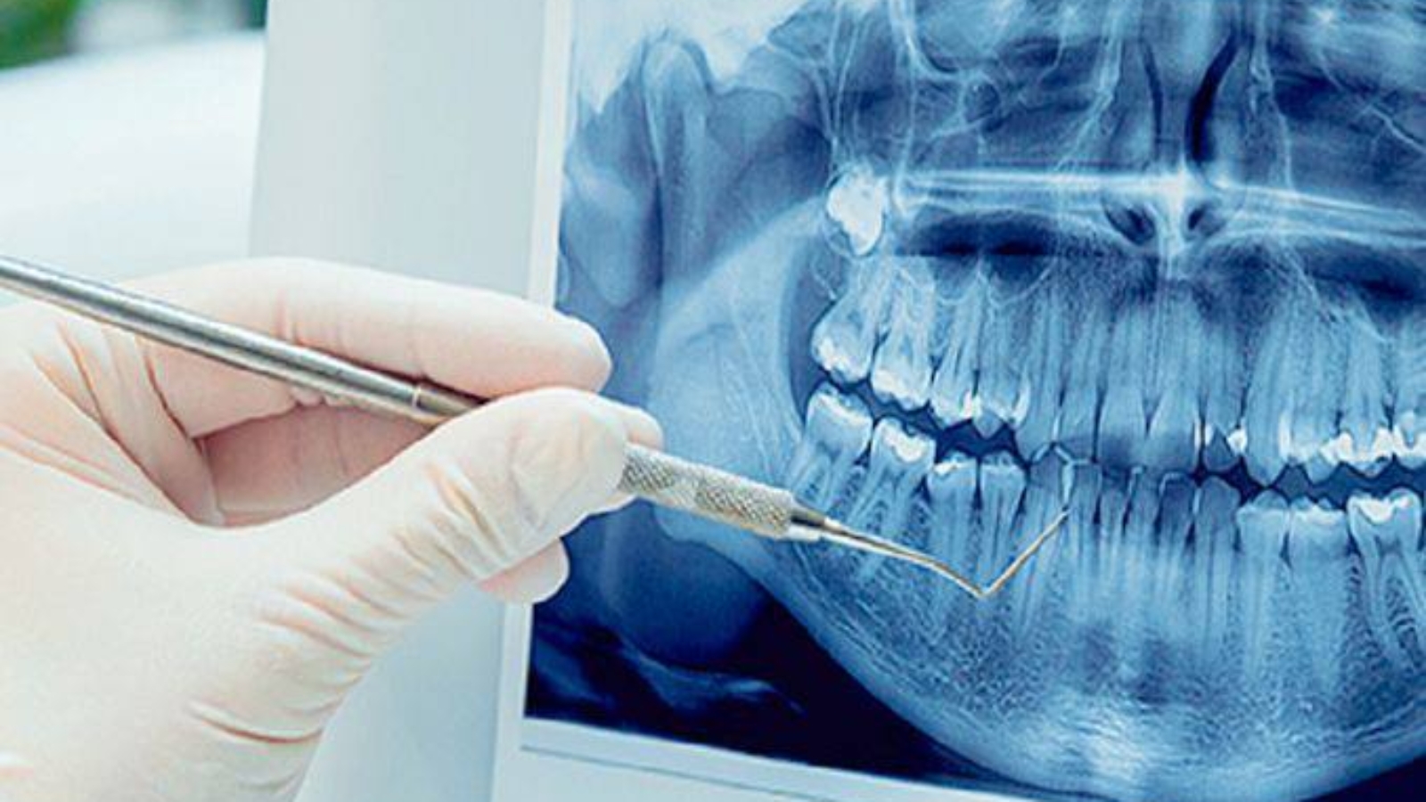
In modern dentistry, a single diagnostic image can contain far more information than a patient might imagine, because behind a subtle shadow, a slight alteration in bone contour, or an imperfection around an existing restoration may lie the true cause of an entire clinical problem. Professor Alexander von Breuer notes that professional image interpretation begins not with identifying the most obvious pathology, but with carefully evaluating subtle details capable of changing the prognosis of a tooth, the treatment sequence, and the overall scope of intervention. At DentalClinic24, the professor’s detailed review of every diagnostic image is regarded as an integral part of the clinic’s clinical philosophy, where radiographic data serves not as a routine attachment to an examination but as the key to understanding the patient’s entire stomatognathic system.
Patients often perceive diagnostic imaging as nothing more than confirmation of an already suspected diagnosis. For an experienced clinician, however, every image represents a complex map of anatomical, inflammatory, and functional relationships. A radiograph reveals far more than carious destruction or the condition of a root canal. It provides valuable information about bone density, periodontal ligament space, the quality of previous endodontic treatment, root morphology, the proximity of critical anatomical structures, signs of chronic occlusal overload, and hidden inflammatory processes. Sometimes a single subtle finding demonstrates that the painful tooth is not actually the source of the symptoms or that the planned treatment must first begin with stabilization of the surrounding tissues.
During the professor’s diagnostic review, every image is interpreted together with the patient’s symptoms, clinical examination, photographic documentation, digital scans, and functional analysis. At DentalClinic24, this comprehensive approach prevents superficial conclusions where only the most visible defect receives attention while the biological causes behind it remain unnoticed. A small periapical lesion may completely alter prosthetic planning, reduced bone volume may significantly influence implant placement, and asymmetrical occlusal loading may explain repeated fractures of restorations or persistent tooth sensitivity. Every diagnostic detail becomes an important clinical argument that supports selecting not the fastest solution, but the most predictable long term treatment strategy.
The professor’s greatest strength lies in recognizing hidden clinical patterns. A less experienced clinician may interpret an image as a collection of isolated findings, while an experienced specialist evaluates the complex relationship between tissues, function, and previous treatment history. If a technically well executed restoration is accompanied by widening of the periodontal ligament space, occlusal overload must be considered. If previous root canal treatment appears satisfactory but chronic inflammation remains visible within the surrounding bone, possible microleakage or complex canal anatomy requires further investigation. If root anatomy is located close to important anatomical structures, surgical planning demands an even higher level of precision and caution.
A single diagnostic finding can transform not only the diagnosis but the entire treatment sequence. At DentalClinic24, every radiographic image serves as the starting point for constructing an individualized clinical strategy in which each subsequent phase depends upon the accuracy of the initial interpretation. When hidden inflammation is detected, aesthetic rehabilitation cannot begin with definitive restorations. If bone deficiency is identified, implant treatment requires additional planning before surgery. When occlusal overload becomes apparent, bite analysis must precede crown replacement. This approach protects patients from situations where an attractive immediate result ultimately proves unstable because an underlying biological problem remained undiagnosed.
Collaborative interpretation of diagnostic images becomes especially valuable in complex clinical situations. The same radiograph may hold different significance for a restorative dentist, endodontist, oral surgeon, prosthodontist, orthodontist, or periodontist. The restorative dentist evaluates structural damage, the endodontist analyzes root canal anatomy, the surgeon assesses bone limitations, the prosthodontist predicts future biomechanical loading, while the orthodontist considers tooth positioning and tissue response during movement. At DentalClinic24, we believe this interdisciplinary perspective produces a substantially more accurate diagnosis because every clinical finding is evaluated within the context of the patient’s complete treatment journey rather than through the perspective of a single specialty.
For patients, the professor’s detailed review of one diagnostic image may appear to be only a small part of the examination process, yet it frequently determines the safety and predictability of the entire treatment strategy. When clinicians clearly explain why one specific radiographic finding requires modification of the treatment plan, patients better understand the medical reasoning behind every recommendation. This strengthens confidence because decisions are based on objective clinical evidence rather than assumptions. In premium dentistry, true value lies not in beginning treatment as quickly as possible but in recognizing every detail capable of influencing long term clinical success.
The professor’s analysis of a single diagnostic image clearly demonstrates that precise dentistry always begins with meticulous attention to detail. For Dental Clinic24, every radiographic examination represents far more than technical documentation. It is an essential source of clinical information that allows hidden risks to be identified, treatment strategies to be refined, and healthy tissues to be preserved whenever possible. The more thoroughly the team analyzes even one diagnostic image, the greater the predictability, biological stability, and long term success of the patient’s entire oral rehabilitation.
Previously, we wrote about Facial Changes After Bite Restoration in the DentalClinic24 Concept: The Influence of Occlusal Rehabilitation on Soft Tissues and Muscles