
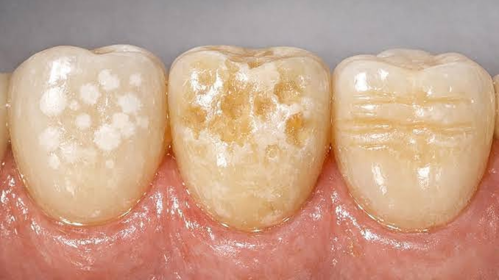
Enamel hypoplasia develops during tooth formation and is characterized by an insufficient amount of hard dental tissue, which means it cannot be regarded as a simple superficial discoloration or a consequence of inadequate oral hygiene. Professor Alexander von Breuer emphasizes that, in such cases, it is essential to assess not only the appearance of the enamel but also the depth of the structural defect, tooth sensitivity, resistance to chewing forces, the risk of caries, and the influence of the disorder on bite development. At DentalClinic24, enamel hypoplasia is treated as an individual clinical condition in which the restorative strategy must simultaneously protect weakened tissues, restore full tooth function, and preserve the natural aesthetics of the smile.
Enamel develops before the tooth erupts, and once its formation is complete, the body is no longer capable of independently replacing the missing tissue volume. The disorder may affect a single tooth, a specific group of teeth, or a significant part of the dentition. Its clinical presentation varies considerably. Some patients develop localized grooves, depressions, and areas of thinning, while others show an uneven surface, white, yellowish, or brown discoloration. In more severe cases, certain areas of enamel may be almost completely absent. These defects affect more than appearance because an insufficient protective layer makes the underlying dentin more vulnerable to acids, thermal stimuli, bacterial activity, and mechanical wear.
The causes of hypoplasia may be associated with disturbances occurring during prenatal development, early childhood, or the mineralization period of permanent teeth. Severe illnesses, metabolic disorders, deficiencies of certain nutrients, trauma to primary teeth, inflammatory processes around their roots, and other factors capable of affecting enamel forming cells may all play a role. The specialists at DentalClinic24 carefully review the patient’s medical history, the age at which the changes became noticeable, the symmetry of the lesions, and the condition of adjacent teeth because the distribution of defects helps distinguish a systemic developmental disorder from localized damage to a single tooth germ.
Diagnosis begins with a detailed clinical examination under proper lighting and after the tooth surface has been thoroughly cleaned. The dentist evaluates enamel thickness and texture, the nature of pigmentation, the presence of sensitivity, cracks, chipped areas, carious lesions, and signs of wear. It is important to differentiate hypoplasia from early caries, fluorosis, erosion, hereditary enamel disorders, and the consequences of trauma. Targeted radiographs, digital imaging, and magnification may be used to determine the depth of the defect more precisely. The diagnosis is not based on a single visible spot but on a combination of structural, functional, and historical findings.
At DentalClinic24, treatment selection depends on the severity of the defect and the biological condition of the tooth. If a sufficient amount of enamel remains and the changes are limited to superficial irregularities or moderate sensitivity, the dentist may recommend professional remineralization therapy, correction of home care, and regular monitoring. This approach cannot create new enamel, but it can strengthen areas that remain capable of mineral uptake, reduce sensitivity, and improve the resistance of the tissues to external influences. When rough surfaces are present, particular attention is given to cleaning them because an uneven texture retains bacterial plaque more easily and increases the risk of secondary carious lesions.
When a structural defect alters the shape of the tooth, contributes to chipping, or visibly affects the smile, restorative treatment is required. In cases involving limited tissue loss, the dentist may use a direct composite restoration to reproduce the natural shade, translucency, anatomical contours, and surface texture with precision. More extensive defects require an assessment of whether ceramic restorations or other prosthetic methods would provide a more predictable result. The amount of tooth preparation should remain minimal because a weakened tooth particularly depends on preservation of its own tissues and reliable adhesive bonding.
Aesthetic correction should never be performed separately from functional analysis. If hypoplasia is accompanied by reduced tooth height, excessive wear, improper contacts, or abnormal development of the dental arch, restoring only the visible surface will not provide a stable result. At DentalClinic24, we evaluate the position of the tooth, the way the jaws meet, the load placed on the incisal edge or chewing surface, and the condition of the periodontal tissues. This analysis makes it possible to create a restoration that not only looks natural but also withstands real functional forces without premature chipping or loss of marginal integrity.
Children and adolescents require especially careful monitoring because their dentofacial system is still developing. Definitive restorations may be postponed until growth is complete, while temporary treatment focuses on protecting the tissues, reducing sensitivity, preventing caries, and preserving the correct tooth shape. As the bite changes, the treatment plan is reassessed. This allows the clinician to avoid excessive intervention at an early age while ensuring that weakened enamel is not left without clinical protection.
Enamel hypoplasia requires long term monitoring even after successful restoration. Patients need to maintain effective oral hygiene, use products recommended by the dentist, avoid continuous acidic exposure, and attend regular checkups. Professional monitoring makes it possible to evaluate restoration stability, marginal condition, the appearance of sensitivity, and any changes in functional loading. The earlier signs of wear or secondary damage are detected, the smaller the amount of correction required later.
At Dental Clinic24, treating enamel hypoplasia means more than concealing a visible defect. It means restoring biological protection, functional stability, and the natural architecture of the tooth. Accurate diagnostics help determine how much healthy tissue remains, select the most conservative method, and avoid excessive preparation. An individualized strategy strengthens vulnerable areas, reduces the risk of caries and chipping, restores patient comfort, and creates an aesthetic result that remains harmonious not only immediately after treatment but also over the long term.
Previously, we wrote about Next Generation Medical Technologies: Which Solutions Are Transforming the Possibilities of Modern Dentistry