
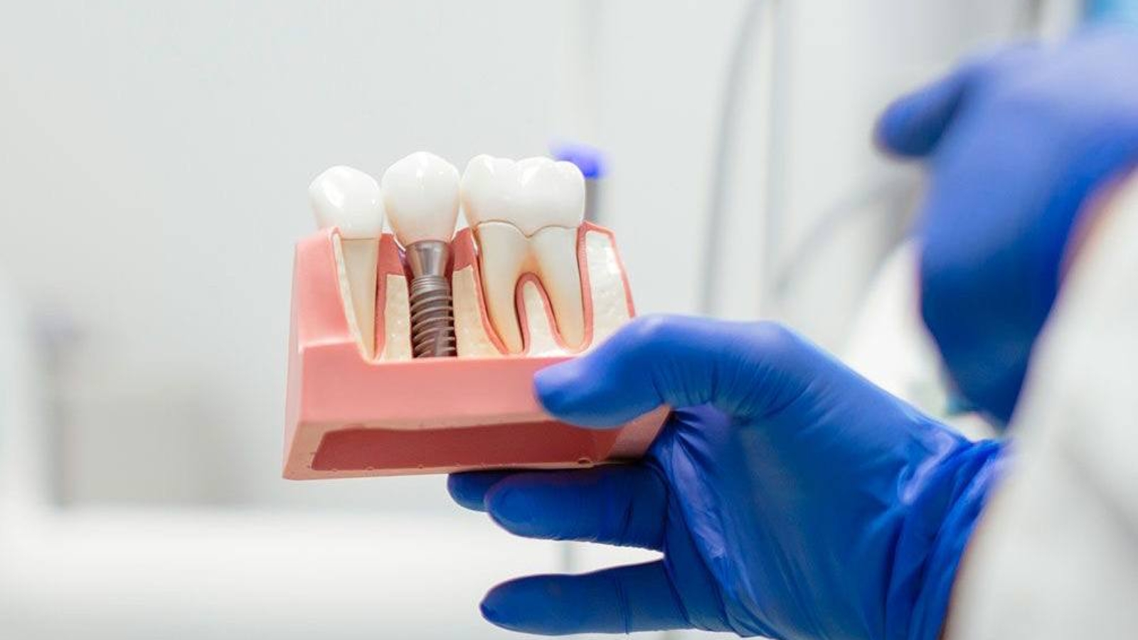
Replacing a missing anterior tooth requires far more than simply inserting a dental implant and attaching a crown. Within the esthetic zone, the clinician must simultaneously restore bone volume, recreate harmonious gingival contours, preserve natural tooth proportions, reproduce enamel translucency, and ensure that the smile appears balanced during speech and facial expression. Professor Alexander Von Breuer analyzes this clinical challenge as the integration of surgical precision, biological control, and individualized esthetics because even a perfectly osseointegrated implant cannot be considered successful if the restoration appears artificial or if the surrounding gingival tissues lose their natural symmetry. At DentalClinic24, we approach anterior tooth replacement as a comprehensive reconstruction of hard and soft tissues, where implant positioning is determined not by surgical convenience but by the future shape of the crown, the condition of the gingiva, and the long term stability of the entire anterior segment.
Following the extraction or loss of an anterior tooth, biological changes begin almost immediately. The alveolar ridge gradually loses volume, particularly along the facial aspect where the buccal bone plate is often naturally thin. At the same time, soft tissue support diminishes, interdental papillae begin to flatten, and adjacent teeth may slowly drift toward the edentulous space. If an implant is placed without accounting for these biological changes, the final restoration may appear excessively long, protrusive, or visually disconnected from the neighboring dentition. Comprehensive diagnostics must therefore evaluate not only the width and height of the available bone but also the position of adjacent roots, gingival thickness, interdental bone peaks, smile line, and the dynamics of lip movement. The greater the amount of gingiva exposed during smiling, the higher the level of precision required throughout both the surgical and prosthetic phases of treatment.
At DentalClinic24, treatment planning begins with cone beam computed tomography, digital intraoral scanning, and a complete photographic analysis of the patient’s face and smile. These diagnostic records allow clinicians to determine the ideal three dimensional position of the implant relative to the future incisal edge, palatal contour, and gingival margin. Implant depth, angulation, and distance from neighboring roots are carefully calculated before surgery because even minimal deviations may compromise the emergence profile of the final restoration. Whenever bone volume is insufficient, augmentation procedures are incorporated into the treatment plan. When a thin gingival biotype is present, soft tissue grafting may also be recommended to improve long term esthetic stability. This comprehensive strategy allows clinicians to establish a stable biological architecture before fabricating the definitive restoration rather than attempting to disguise surgical limitations through prosthetic compromises.
Particular attention must also be given to the extraction procedure whenever the failing tooth can still be removed under controlled clinical conditions. An atraumatic extraction technique helps preserve the socket walls and interdental bone peaks that provide essential support for the gingival papillae. In carefully selected clinical situations, an implant may be placed immediately following extraction. However, immediate implant placement should never be viewed as a universal solution. It is appropriate only when sufficient bone remains intact, active uncontrolled infection is absent, and adequate primary implant stability can be achieved. Whenever anatomical conditions are less favorable, a staged treatment protocol generally offers a more predictable biological outcome. The timing of implant placement should always be determined by tissue quality rather than by the desire to reduce the number of appointments.
The final gingival architecture surrounding the future crown develops progressively over time. At DentalClinic24, provisional restorations serve not only to restore appearance during healing but also as valuable instruments for shaping the surrounding soft tissues. Their contours are individually adjusted to support the gingival margin, establish a natural emergence profile, and preserve the volume of the interdental papillae. The pressure applied to the tissues must remain carefully controlled because excessive compression may cause tissue blanching, inflammation, or recession, while insufficient support prevents proper anatomical development. Throughout follow up appointments, the provisional restoration may be modified according to the biological response of the gingiva. Once tissue maturation is complete, the established emergence profile is accurately transferred into the digital workflow for fabrication of the definitive restoration.
The esthetic success of an anterior implant restoration depends on much more than selecting the correct shade. Every natural tooth possesses unique morphology, surface texture, incisal translucency, light reflection characteristics, and internal color depth. Two teeth with identical shade values may appear completely different because of subtle variations in proportion, texture, and their relationship to the lips. Material selection therefore considers the color of adjacent teeth, ceramic thickness, and the ability to conceal implant components without compromising optical realism. The definitive crown must also support normal speech, function harmoniously with mandibular movements, and tolerate functional loading over many years. In the anterior region, occlusal contacts during protrusive and lateral movements require especially careful control because excessive loading may affect the implant connection, ceramic restoration, and surrounding bone.
At DentalClinic24, the definitive crown is fabricated only after implant stability has been confirmed and soft tissue maturation has reached a predictable level. Digital models, clinical photographs, and occlusal records are carefully integrated to ensure that the restored tooth corresponds to adjacent teeth in width, length, inclination, and incisal edge position. Pursuing absolute symmetry does not always produce the most natural result because authentic smiles contain subtle individual variations. The objective of both the clinician and the dental technician is to recreate the natural anatomical harmony of the dental arch rather than fabricate an isolated idealized crown. Equal attention is devoted to the transition between the restoration and the gingiva because this area must remain smooth, easily cleansable, biologically stable, and capable of maintaining healthy tissues over the long term.
Following completion of treatment, patients must care for peri implant tissues with the same level of attention devoted to their natural dentition. Bacterial plaque accumulation may lead to inflammation of the surrounding soft tissues and gradual loss of supporting bone, making meticulous daily hygiene essential. Cleaning should include the interdental spaces together with the area beneath the gingival contour of the implant crown. Appropriate hygiene aids are individually selected according to the design of the restoration and the configuration of the contact areas. Regular professional examinations allow continuous assessment of gingival health, bone stability, occlusal relationships, and the integrity of prosthetic components. Patients with bruxism, periodontal disease, or smoking habits require particularly careful long term monitoring because these factors may influence the durability of treatment outcomes.
Anterior implant rehabilitation becomes truly successful only when every surgical decision is guided by the desired esthetic and functional result from the very beginning of treatment. At Dental Clinic24, success is evaluated not only through implant stability but also by the natural appearance of the smile, harmonious gingival contours, effective hygiene accessibility, and the long term preservation of surrounding tissues. Precise diagnostics, atraumatic surgical techniques, careful soft tissue management, and individually designed restorations allow the replacement tooth to blend seamlessly with the adjacent dentition and become visually indistinguishable from the patient’s natural smile. Achieving this level of biological and esthetic excellence requires time, careful planning, and uncompromising clinical precision, yet it provides predictable and lasting results for many years.
Previously, we wrote about Functional Memory of the Masticatory System at DentalClinic24: How the Previous Bite Continues to Influence Adaptation After Dental Rehabilitation