
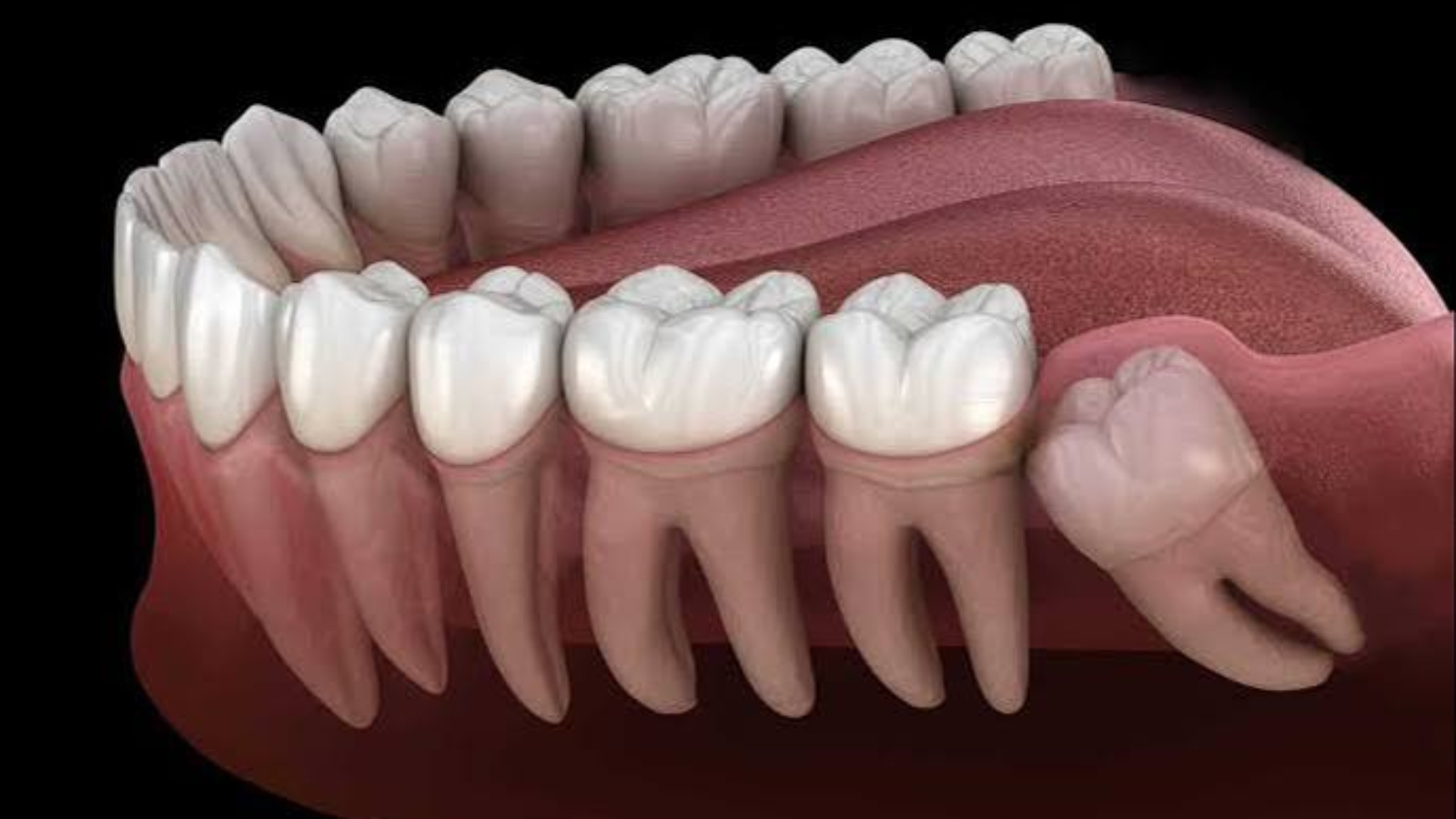
Wisdom teeth are surrounded by numerous misconceptions that often cause patients either to insist on extraction without sufficient clinical justification or to postpone examination until significant pain and inflammation develop. Third molars differ from other teeth not because of unique biological characteristics but because of their late eruption, highly variable anatomical position, and the limited amount of available space within the posterior regions of the jaws. Professor Alexander Von Breuer believes that every clinical decision should be based not simply on the presence of a wisdom tooth but on its anatomical position, the condition of the surrounding tissues, its relationship with neighboring teeth, and its long term prognosis. At DentalClinic24, we evaluate every third molar individually because a fully erupted and functional wisdom tooth may remain healthy for decades, while an impacted tooth without symptoms may already be creating risks for adjacent teeth, surrounding bone, or soft tissues.
One of the most widespread misconceptions is that every wisdom tooth should be removed as a preventive measure. This assumption does not reflect modern clinical principles. When a third molar erupts vertically, participates effectively in chewing, can be cleaned properly, and does not traumatize surrounding tissues or create pathological pressure on neighboring structures, surgical removal may not be necessary. Conversely, the absence of symptoms does not always indicate the absence of disease. An impacted wisdom tooth may remain completely enclosed within bone, while a partially erupted tooth may be covered by a gingival flap that gradually traps bacterial plaque. Pathological changes in this region frequently develop slowly and remain unnoticed for extended periods. Accurate diagnosis therefore requires both a comprehensive clinical examination and appropriate radiographic imaging rather than relying solely on the patient’s subjective sensations.
At DentalClinic24, particular attention is devoted to evaluating the relationship between the wisdom tooth and the adjacent second molar. When the third molar is positioned horizontally or at a significant angle, its crown may press directly against the distal surface of the neighboring tooth, creating an area that becomes extremely difficult to clean adequately. Such anatomical conditions substantially increase the risk of proximal caries, gingival inflammation, and gradual destruction of the supporting alveolar bone. Damage affecting the second molar often has greater long term functional consequences than preserving the wisdom tooth itself. Clinical decisions are therefore based on protecting healthy neighboring teeth whenever predictable complications can be anticipated. Cone beam computed tomography provides significantly more detailed information regarding root orientation, surrounding bone anatomy, and the spatial relationship between adjacent structures than conventional visual examination alone.
Pain associated with a wisdom tooth is not always caused by the eruption process itself. The underlying cause may involve inflammation of the surrounding soft tissues, dental caries, excessive occlusal loading, trauma produced by the opposing tooth, or infection spreading into neighboring anatomical spaces. During pericoronitis, the gingival tissue covering a partially erupted third molar becomes swollen, painful, and difficult to clean effectively. Patients may additionally experience unpleasant taste, restricted mouth opening, and discomfort while swallowing. Temporary reduction of symptoms does not eliminate the anatomical cause responsible for recurrent inflammation. The clinician must determine whether favorable conditions for complete eruption and adequate oral hygiene can realistically be achieved or whether preserving the tooth will continue to promote repeated inflammatory episodes.
Another common misconception concerns the belief that wisdom teeth inevitably cause crowding of the anterior teeth. Changes in the position of the incisors frequently occur during the same period in life when third molars erupt, but this coincidence alone does not establish a direct causal relationship. Long term stability of the dental arch is influenced by jaw growth, periodontal health, occlusal relationships, muscular forces from the lips and tongue, and the absence of proper retention following orthodontic treatment. At DentalClinic24, we do not regard wisdom tooth extraction as a universal method for preventing crowding of the anterior teeth. Surgical removal should always be supported by independent clinical indications, while long term orthodontic stability depends primarily on appropriate retention protocols and continuous monitoring of functional factors.
The complexity of wisdom tooth extraction depends far more on anatomical characteristics than on crown size or patient age alone. The depth of impaction, root morphology, bone density, proximity to the inferior alveolar nerve, relationship to the maxillary sinus, and the presence of inflammatory changes all significantly influence surgical planning. In the lower jaw, third molar roots may develop very close to the nerve responsible for sensation of the lower lip and chin. In the upper jaw, the floor of the maxillary sinus becomes an equally important anatomical landmark. Three dimensional imaging allows clinicians to evaluate these relationships before surgery, determine the safest surgical approach, and decide whether sectioning the tooth into several components will allow more conservative and controlled removal.
Wisdom tooth extraction should never become an unnecessarily traumatic procedure. At DentalClinic24, every surgical protocol is designed to preserve surrounding bone, protect the adjacent second molar, and minimize injury to the surrounding soft tissues. Following effective anesthesia, the surgeon carefully gains access to the tooth, divides the crown and roots whenever clinically indicated, removes each component in a controlled manner, and thoroughly cleans the surgical site. This approach minimizes excessive force upon the jaw while allowing complete control throughout the extraction process. The duration of surgery depends primarily on anatomical complexity rather than technical quality. More important than procedural speed are surgical precision, effective hemostasis, and meticulous closure of the surgical wound.
Following extraction, patients may experience swelling, moderate discomfort, and temporary limitation of mouth opening, particularly when deeply impacted wisdom teeth have been removed. These postoperative reactions represent the body’s normal biological response to surgery and generally decrease gradually during healing. Patients should carefully follow postoperative instructions, avoid disturbing the extraction site, maintain appropriate oral hygiene, and take prescribed medications exactly as directed. Increasing pain after an initial period of improvement, persistent unpleasant odor, elevated body temperature, ongoing bleeding, or progressive numbness require immediate professional evaluation. Timely postoperative assessment allows clinicians to distinguish normal healing from conditions requiring additional treatment or modification of postoperative management.
At Dental Clinic24, our primary objective is never the extraction or preservation of a wisdom tooth as an isolated goal but rather the long term protection of the entire dentofacial system from predictable complications. A third molar may remain a healthy and functional component of the dentition whenever its position, surrounding tissues, and accessibility for hygiene permit favorable long term prognosis. However, when risks involving the adjacent tooth, recurrent inflammation, cyst formation, bone destruction, or other clinically confirmed pathological changes are present, timely extraction becomes a justified preventive measure. Every decision should be guided by accurate diagnostics rather than myths, age related stereotypes, or waiting until severe pain develops. This evidence based approach preserves healthy tissues while ensuring that surgical intervention, when necessary, is performed under the safest and most predictable clinical conditions.
Previously, we wrote about Orthopantomogram in Diagnostics at DentalClinic24: The Value of Comprehensive Imaging for Precise Treatment Planning