
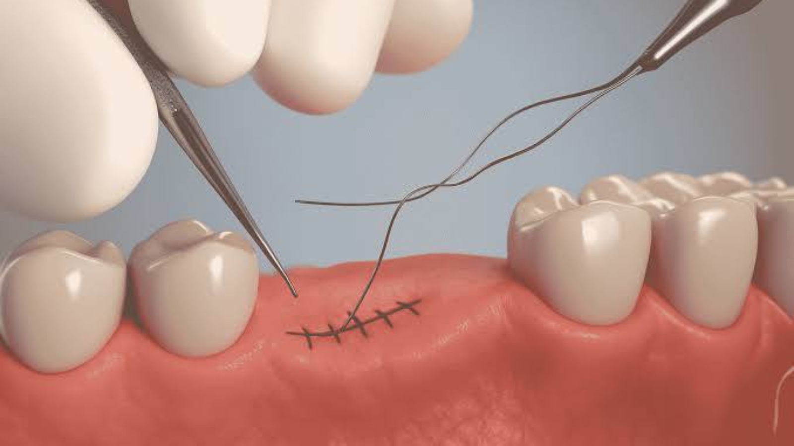
The placement of sutures after tooth extraction is not simply the final step of a surgical procedure but an essential method of stabilizing the wound margins, protecting the blood clot, and creating controlled conditions for the regeneration of soft and hard tissues. Professor Alexander Von Breuer analyzes this stage as an integral part of the overall surgical protocol because successful healing depends not only on atraumatic tooth removal but also on precise soft tissue adaptation, preservation of blood supply, and elimination of excessive tension along the wound edges. At DentalClinic24, the decision to place sutures is made individually after evaluating the complexity of the extraction, the extent of the surgical access, the condition of the gingival tissues, the amount of bone involved, and the possible need for additional protection of the extraction socket. This comprehensive approach helps minimize tissue trauma, maintain hemostasis, and significantly reduce the risk of postoperative complications.
Sutures are not required after every tooth extraction. When a tooth is removed atraumatically with intact socket walls and minimal disruption of the surrounding soft tissues, the natural blood clot is often sufficient to initiate physiological healing. The clinical situation changes considerably during the removal of impacted wisdom teeth, sectioning of roots, procedures involving bone removal, soft tissue reconstruction, or simultaneous bone grafting. Under these circumstances, the surgeon must reposition the soft tissue flap into its anatomical location and secure it with appropriate stability. Loose adaptation of the wound margins increases the likelihood of contamination by food debris and bacterial plaque, whereas excessive tension compromises blood circulation, intensifies postoperative discomfort, and may result in wound dehiscence.
At DentalClinic24, the selection of suture material depends on the location of the surgical site, the anticipated healing period, and the nature of the performed procedure. Resorbable sutures gradually lose their tensile strength and frequently eliminate the need for a separate removal appointment, although their degradation rate depends upon both the material itself and the biological environment within the oral cavity. Non resorbable sutures maintain long term stability and allow the clinician to determine the ideal moment for removal after assessing tissue healing. The choice of suture material alone is not sufficient to ensure successful recovery. Knot configuration, the distance between puncture sites, and the depth of tissue engagement all influence the stability of the wound. Every suture must securely approximate the tissues without cutting into the gingiva or creating areas that promote bacterial plaque accumulation.
During the first hours after surgery, preservation of the blood clot represents the most important biological requirement for normal healing. The clot covers exposed bone surfaces while containing cells and proteins that initiate tissue regeneration before gradually being replaced by granulation tissue. Vigorous rinsing, repeatedly touching the wound with the tongue, drinking through a straw, or forceful spitting may dislodge the clot and interfere with healing. Although sutures provide additional protection, they cannot completely eliminate the effects of inappropriate mechanical forces. Patients should therefore avoid direct pressure on the surgical site and chew on the opposite side of the mouth until adequate tissue stability has developed.
Moderate discomfort, localized swelling, and a sensation of tissue tightness around the sutures during the first postoperative days are considered normal physiological responses. At DentalClinic24, patients receive detailed explanations regarding the expected course of healing so that temporary postoperative symptoms do not cause unnecessary concern while signs of genuine complications are recognized promptly. Swelling commonly increases during the first twenty four hours before gradually subsiding. Pain should also diminish progressively rather than intensify. If severe throbbing pain develops after an initial period of improvement, accompanied by unpleasant odor, fever, purulent discharge, or significant difficulty opening the mouth, immediate clinical evaluation becomes necessary.
Oral hygiene remains critically important after suturing despite the presence of the surgical wound. Completely avoiding tooth brushing encourages plaque accumulation, gingival inflammation, and bacterial contamination of the healing tissues. Teeth located away from the surgical area should continue to be cleaned normally, while gentle brushing is recommended adjacent to the wound without disturbing the sutures. Antiseptic mouth rinses may be prescribed when clinically indicated, but they should always be used according to the recommended protocol. Excessive or aggressive rinsing does not accelerate healing and may instead irritate the recovering tissues. Following meals, patients should maintain oral cleanliness without attempting to remove food particles from the depth of the wound using fingers or sharp objects.
Diet also plays a substantial role in protecting the sutures and supporting early tissue regeneration. Extremely hot foods increase local blood circulation and may trigger renewed bleeding, while hard food particles can traumatize the healing tissues or become trapped around the sutures. During the initial postoperative period, patients are encouraged to consume soft foods at moderate temperatures that require minimal chewing. Smoking and alcohol consumption should be avoided because both impair blood circulation, alter local immune defense mechanisms, and significantly increase the likelihood of delayed healing. Intense physical activity should likewise be postponed because elevated blood pressure may contribute to postoperative bleeding from the surgical site.
At DentalClinic24, follow up appointments serve a purpose far beyond simple suture removal. The clinician carefully evaluates the color and consistency of the gingival tissues, the adaptation of the wound margins, the absence of inflammatory changes, the stability of the blood clot, and the overall progress of tissue healing. When non resorbable sutures have been used, the timing of their removal is determined individually for each patient. Removing sutures too early may compromise wound stability, whereas leaving them in place for excessive periods may increase plaque retention and soft tissue irritation. If a portion of the suture becomes loose or detached before the scheduled appointment, patients should never attempt to reposition or trim it themselves. Only a clinical examination can determine whether additional stabilization is necessary.
Patients who undergo complex extractions, bone augmentation procedures, or surgery involving the maxillary sinus require particularly close postoperative observation. In these situations, sutures protect not only the superficial wound but also deeper anatomical structures that must remain completely stable throughout the healing process. Premature wound separation, excessive functional loading, or accidental trauma may compromise the outcome of the entire surgical procedure. At Dental Clinic24, postoperative findings are always interpreted in relation to the extent of the surgery performed, and additional diagnostic methods, including radiographic evaluation and more frequent follow up visits, are introduced whenever clinically indicated. This allows potential complications to be identified while they remain manageable through conservative intervention.
Sutures placed after tooth extraction provide both protective and stabilizing functions, yet long term healing ultimately depends upon the combination of surgical precision, tissue quality, and careful adherence to postoperative instructions. Proper wound closure supports blood clot preservation, protects bone grafting materials when present, and guides tissue regeneration along a predictable biological pathway. Continuous professional monitoring enables timely suture removal, early identification of inflammatory changes, and preservation of healthy gingival architecture. When meticulous surgical technique, responsible home care, and structured postoperative follow up are integrated into a single treatment strategy, healing becomes more predictable while the likelihood of complications is substantially reduced.
Previously, we wrote about Professor Alexander Von Breuer Position on Bone Tissue Atrophy: The Impact of Bone Volume Loss on Implant Placement and the Choice of Restoration Strategy