
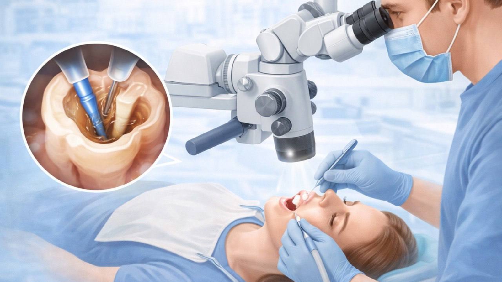
A dental microscope is not simply a tool that magnifies the operating field. It is an instrument that allows clinicians to obtain diagnostic information that cannot be evaluated reliably with the naked eye. Professor Alexander Von Breuer analyzes optical magnification as one of the most important technologies supporting controlled and minimally invasive dentistry because microcracks, accessory root canals, concealed areas of softened dentin, and imperfections at restoration margins often measure only fractions of a millimeter. At DentalClinic24, we integrate the dental microscope into every clinically significant stage of treatment. This approach enables us to distinguish diseased tissue from healthy tooth structure, accurately determine the true boundaries of defects, and perform treatment without unnecessarily sacrificing intact dental tissues.
The greatest advantage of magnification lies in the combination of superior optics and concentrated illumination. During conventional examination, deep portions of a cavity often remain partially shadowed, while subtle color differences between healthy and altered dentin may be difficult to distinguish. A dental microscope creates an intensely illuminated and highly magnified working field that allows clinicians to evaluate tissue relief, crack orientation, restoration margins, and microscopic surface changes in enamel with exceptional precision. Variable magnification levels are equally important. Lower magnification provides an overall anatomical perspective, medium magnification supports cavity preparation and restorative procedures, while high magnification is indispensable when working within narrow root canals, identifying microperforations, or evaluating delicate interfaces between restorative materials and natural tissues.
At DentalClinic24, microscopic diagnostics begin before active treatment starts. We correlate the patient’s symptoms with radiographic findings, clinical testing, and microscopic visualization. If discomfort occurs during chewing or exposure to temperature changes, the underlying cause may extend beyond conventional caries and include hidden enamel cracks, defective restoration margins, localized dentin thinning, or pulpal inflammation. Under magnification, clinicians can identify subtle color variations surrounding crack lines, determine their direction, and evaluate whether the defect remains confined to enamel or extends into deeper structures. This distinction has a direct impact on prognosis because superficial enamel fractures and vertical root fractures require fundamentally different therapeutic approaches.
During caries treatment, microscopic visualization makes it possible to precisely control the amount of tissue being removed. Infected softened dentin must be eliminated, yet excessive preparation weakens the remaining tooth structure and reduces its ability to withstand functional loading. Without sufficient visual control, clinicians often rely primarily on tactile sensation and overall appearance. Magnification allows gradual evaluation of tissue quality in highly localized areas throughout the preparation process. Healthy, structurally sound dentin can therefore be preserved wherever removal offers no biological advantage, while narrow regions along cavity margins that frequently harbor bacterial contamination can be thoroughly cleaned. This selective approach becomes especially valuable when treating deep lesions located close to the dental pulp.
Endodontic treatment represents one of the clinical disciplines in which optical magnification has a direct influence on tooth preservation. Root canal systems rarely consist of straight and uniformly positioned canals. Accessory canals, isthmuses, calcified canal entrances, and complex anatomical curvatures are frequently encountered within the same tooth. At DentalClinic24, microscopic visualization allows clinicians to locate canal orifices without unnecessarily removing dentin from the chamber floor. Natural anatomical landmarks, subtle color transitions, and microscopic surface details clearly indicate canal locations. This significantly reduces the risk of overlooking infected anatomy, creating false pathways, or preparing excessively large access cavities that compromise the structural integrity of the remaining tooth.
Retreatment of previously treated root canals demands an even greater level of visual precision. Existing filling materials, separated instruments, ledges, and alterations created during earlier procedures often complicate access to the apical portion of the canal system. Under magnification, clinicians can gradually separate filling material from canal walls, continuously evaluate dentin thickness, and carefully reestablish the original canal pathway. When a fractured instrument is detected, the decision to remove it depends upon its location, the thickness of surrounding dentin, and the potential risk of perforation. Preserving a tooth should never come at the expense of dangerously weakening the root. The microscope therefore serves not only as a treatment aid but also as a guide for determining the safest biological limits of intervention.
Magnification also substantially improves the quality of restorative dentistry by allowing meticulous adaptation of composite materials and careful control of every restorative layer. Restoration margins must transition seamlessly into enamel without steps, overhangs, or microscopic gaps capable of accumulating bacterial plaque. At DentalClinic24, we examine cavity margins, isolation quality, composite adaptation, and contact point morphology under magnification. Particular attention is given to subgingival areas and interdental surfaces because these regions are difficult to evaluate directly and often present the greatest challenges for daily oral hygiene. The more accurately restorative materials integrate with natural tooth tissues, the lower the probability of recurrent caries, gingival inflammation, and premature restoration replacement.
Microscope assisted dentistry requires considerably more than advanced optical equipment. Every component of the clinical workflow must support precision. The clinician works in a stable ergonomic position while utilizing rubber dam isolation, microsurgical instruments, and highly efficient aspiration systems. Absolute isolation is especially important during adhesive procedures because even minimal contamination with saliva may compromise the bond between restorative materials and enamel or dentin. Rubber dam isolation simultaneously protects the working field, surrounding soft tissues, and the patient’s airway. Under magnification, clinicians can monitor isolation quality continuously, identify any potential leakage, and maintain a completely dry and well illuminated treatment environment throughout every stage of the procedure.
Microscopic documentation represents another important advantage of modern dental treatment. High resolution photographs and video recordings preserve the initial clinical condition, identified defects, treatment stages, and final results. These records allow clinicians to compare findings over time, explain complex clinical situations to patients, and maintain objective documentation of completed procedures. In complicated cases, recordings may also facilitate collaborative evaluation by several specialists. This approach becomes particularly valuable when determining whether a tooth should undergo restorative treatment, prosthetic reinforcement, or extraction. Clinical decisions are based on directly visualized evidence rather than subjective assumptions.
Magnification also plays an important role during tooth preparation for veneers, ceramic inlays, and crowns. Tooth preparation must create adequate space for restorative materials while preserving as much healthy tissue as biologically possible. A microscope enables clinicians to control preparation depth, refine surface morphology, and establish precise finish lines that dental technicians can reproduce accurately. If the preparation margin is irregular or extends excessively below the gingival tissues, both conventional impressions and digital scans may lose accuracy, reducing the precision of the definitive restoration. At DentalClinic24, optical magnification allows us to preserve enamel wherever possible while creating preparation geometry that ensures predictable long term retention and restoration stability.
We regard the dental microscope not as an isolated technology but as a clinical philosophy that increases the precision and biological justification of every treatment decision. Magnification allows earlier detection of microscopic defects, more accurate assessment of lesion depth, preservation of greater volumes of healthy tooth structure, and consistent quality control in anatomically challenging areas. At Dental Clinic24, the true value of microscope assisted dentistry lies in its ability to make treatment more selective, conservative, and predictable. The more precisely clinicians can visualize tooth anatomy, the less frequently they need to remove healthy tissues as a safety margin, resulting in better preservation of natural tooth strength, function, and long term structural integrity.
Previously, we wrote about The Anatomical Limits of Tooth Restoration at DentalClinic24: Where the Boundary Lies Between Treatment and Overload