
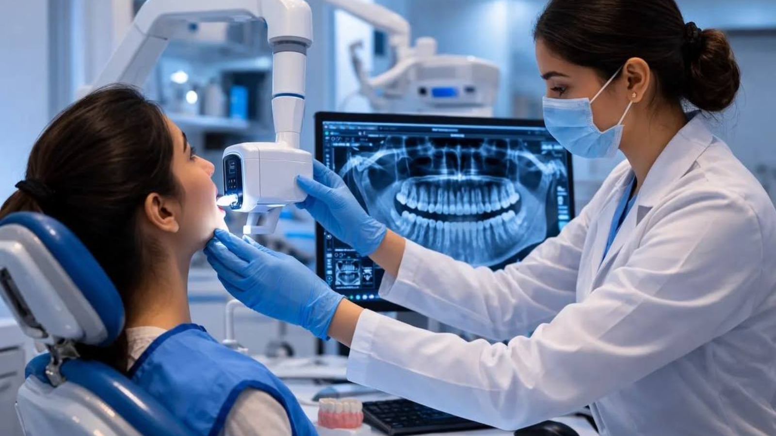
Technological precision in dentistry does not begin with purchasing new equipment. It begins with understanding which clinical challenge a particular technology is intended to solve and how the information it provides will influence treatment for an individual patient. Professor Alexander Von Breuer sees this as one of the defining principles of modern dentistry because technology delivers genuine value only when it is fully integrated into diagnosis, treatment planning, and long term clinical evaluation. At DentalClinic24, we assess every technological innovation according to its ability to reduce clinical uncertainty, preserve healthy biological tissues, and make treatment outcomes more predictable. Our priority is never the complexity of the equipment itself or the number of digital features it offers, but rather the accuracy of its measurements, the consistency of its results, patient safety, and its contribution to every stage of the clinical workflow.
One of the most significant advances in contemporary dentistry is three dimensional imaging through cone beam computed tomography. Conventional radiographs display anatomical structures in only one plane and may conceal overlapping roots, bone defects, or important anatomical landmarks. Three dimensional imaging allows clinicians to evaluate root canal morphology, the position of the inferior alveolar nerve, available bone volume, the condition of the maxillary sinuses, the location of impacted teeth, and hidden inflammatory lesions with far greater precision. Nevertheless, the true value of tomography lies not in producing detailed images but in the professional interpretation of those findings. Every scan must be correlated with the clinical examination, patient symptoms, soft tissue evaluation, and functional assessment because digital imaging supports clinical reasoning rather than replacing it.
At DentalClinic24, three dimensional diagnostic information serves as the foundation for precise surgical planning. Before implant placement, we evaluate not only available bone volume but also the ideal position of the future prosthetic crown, anticipated functional loading, soft tissue thickness, and the relationship between implants and neighboring anatomical structures. Based on these digital models, surgical guides may be fabricated to define the exact direction and depth of implant site preparation. This approach becomes especially valuable when bone volume is limited, multiple implants are required, or complete arch rehabilitation is planned. Guided surgery allows virtual treatment planning to be transferred into the clinical environment with exceptional precision. However, final surgical decisions always depend upon the actual biological condition of the bone, primary implant stability, and tissue response observed during the procedure.
Digital intraoral scanning has transformed many aspects of restorative and prosthetic dentistry by gradually replacing conventional impression materials. Modern scanners accurately capture tooth morphology, gingival contours, implant positions, and occlusal relationships while producing highly detailed digital models. These models enable clinicians to enlarge specific areas, verify preparation margins, and transmit information directly to the dental laboratory without the dimensional changes that may occasionally occur during transportation of conventional impressions. Patients generally experience greater comfort during digital scanning, particularly those with a pronounced gag reflex or limited mouth opening. Clinicians also benefit from the ability to immediately identify and rescan isolated areas requiring greater accuracy without repeating the entire impression procedure.
High precision becomes particularly important when manufacturing ceramic inlays, veneers, crowns, and implant supported restorations. Even minimal inaccuracies at restoration margins may compromise sealing, encourage plaque accumulation, or negatively influence soft tissue stability. A completely digital workflow allows intraoral scans, clinical photographs, tomographic data, and restorative designs to be combined into one comprehensive treatment model. Dental technicians receive detailed digital information rather than an isolated physical impression, allowing them to evaluate proximal contacts, restorative thickness, and available prosthetic space before fabrication begins. This process does not eliminate manual craftsmanship or professional judgment but significantly expands quality control opportunities before definitive restorations are produced.
At DentalClinic24, the operating microscope is regarded as an essential clinical instrument rather than an optional technological addition. Magnification combined with focused illumination enables clinicians to identify microscopic fractures, additional root canals, residual restorative materials, hidden carious lesions, and imperfections along restoration margins that would otherwise remain undetected. In endodontics, the microscope provides visual access to areas deep within the tooth where conventional visibility is extremely limited. During retreatment procedures, clinicians can more accurately remove previous filling materials, locate untreated anatomical structures, and preserve a greater amount of healthy dentin. In restorative dentistry, enhanced visualization allows cavity preparation margins to be refined with greater precision while ensuring optimal adaptation between restorative materials and natural enamel. The better clinicians visualize their working field, the less unnecessary removal of healthy tissue becomes necessary.
The precision of surgical procedures is further enhanced through piezosurgical technology. Using ultrasonic micro vibrations specifically designed for bone surgery, these systems enable highly controlled preparation of mineralized tissues while reducing the likelihood of damaging adjacent soft anatomical structures during selected surgical procedures. Piezosurgery is particularly valuable for complex tooth extractions, implant site preparation, bone grafting, and procedures performed near delicate anatomical regions. Nevertheless, instrument selection always depends upon the individual clinical situation, bone density, surgical objectives, and anatomical conditions. No technological system represents a universal solution, making it essential for clinicians to understand both the advantages and the limitations of every instrument they employ.
Another important technological direction involves controlling microbial contamination. Modern irrigation systems, ultrasonic activation of disinfecting solutions, and photodynamic therapy improve the decontamination of anatomical areas that conventional mechanical instrumentation cannot completely reach. Within root canal systems, bacteria frequently persist inside lateral canals and dentinal tubules, while periodontal biofilms may remain deeply embedded beneath the gingival margin. Additional activation techniques enhance antimicrobial effectiveness, although they cannot compensate for diagnostic errors or incomplete removal of infected tissues. We consider these technologies integral components of comprehensive treatment protocols in which every stage strengthens the biological success of the previous one.
At DentalClinic24, introducing new equipment is always accompanied by comprehensive professional training and continuous refinement of clinical protocols. Installing advanced technology alone is never sufficient because clinicians must understand its physical principles, operating parameters, potential limitations, and quality control standards. Dental assistants receive specialized instruction in equipment preparation, component maintenance, and efficient organization of the clinical environment. Specialists routinely analyze completed cases, compare digital treatment plans with actual clinical outcomes, and continuously optimize procedural workflows. This systematic approach ensures that technology is applied thoughtfully and consistently while making precision the result of coordinated teamwork rather than the presence of sophisticated equipment alone.
Modern digital systems also improve long term treatment monitoring after active care has been completed. Digital models can be compared over time to evaluate tooth wear, gingival changes, tooth movement, and the stability of prosthetic restorations. Follow up radiographic examinations allow clinicians to monitor bone healing, assess periapical tissues, and verify implant integration. Standardized photographic documentation records changes in tooth color, morphology, and soft tissue architecture, which is particularly valuable during aesthetic rehabilitation. The accumulation of objective clinical data allows treatment outcomes to be evaluated not only according to patient perception but also through measurable biological and functional parameters.
Technological progress in dentistry should never increase the number of procedures performed without genuine clinical indications. Its true purpose is to make treatment more precise, less invasive, safer, and biologically more predictable. At Dental Clinic24, we implement new technologies only when they improve diagnostic accuracy, enhance treatment planning, reduce surgical trauma, or provide objective verification of clinical outcomes. The strength of an advanced dental clinic is determined not by the quantity of equipment it possesses but by its ability to integrate technological innovation with clinical expertise, biological principles, and responsible professional judgment. When digital information is interpreted by experienced clinicians, technology becomes far more than a demonstration of modern equipment. It becomes a reliable instrument for achieving long term dental health and predictable treatment success.
Previously, we wrote about Trismus in the Dental Practice of DentalClinic24: Causes of Limited Mouth Opening, Diagnosis, and Treatment Strategy