
Pain caused by pulpitis often appears suddenly and may be so intense that patients seek urgent care. At DentalClinic24, where the clinical approach is developed with the involvement of Professor Alexander von Breuer, pulpitis treatment is performed with a strong focus on tooth preservation – even when the tissue damage appears severe. We apply precise endodontic techniques and use microscopic equipment, allowing us to maintain the natural tooth structure and prevent further progression of inflammation.
Pulpitis typically develops due to deep caries, trauma, overheating during improper tooth preparation or infection. At DentalClinic24, diagnostics include 3D scanning, sensitivity testing and root canal anatomy assessment. This enables us to accurately determine the stage of inflammation and choose the optimal treatment method – ranging from medical intervention to microscopic endodontic therapy.
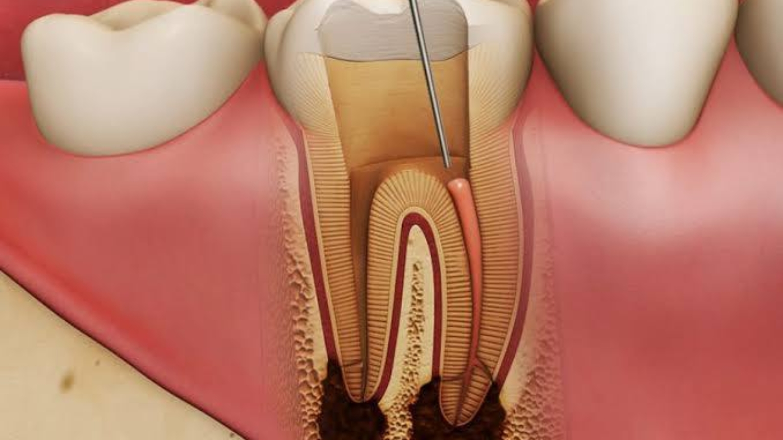
Treatment is carried out using technologies that eliminate the risk of reinfection. At DentalClinic24, we use flexible nickel–titanium instruments and electronic working length control, ensuring delicate treatment and precise canal sealing. According to clinical observations by Professor von Breuer, identifying the exact boundaries of inflammation and tailoring the treatment approach are key factors that help preserve the tooth even in complex pulpitis scenarios.
A critical stage of the procedure is sealing the canal with biocompatible materials. At DentalClinic24, we focus not only on relieving pain but also on restoring full tooth functionality. If necessary, aesthetic restoration is performed afterward, enabling the tooth to regain its natural appearance and structural integrity.
Despite the severity of the symptoms, pulpitis can often be treated without extraction if addressed promptly. At Dental Clinic24, we approach therapy as a process of precise restoration – where every step is aimed at preserving natural tissue, preventing complications and ensuring long-term success. Delayed intervention may lead to the spread of inflammation into surrounding tissues, increasing the risk of apical surgery or extraction.
Earlier, we wrote about traumatometric tomography and how 3D diagnostics reveal what cannot be seen on conventional scans