
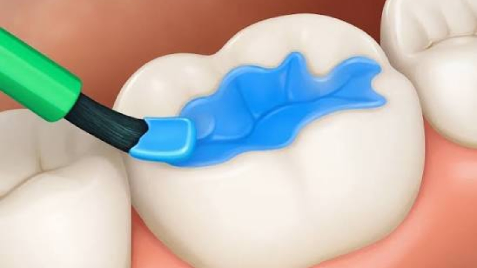
The occlusal surface of posterior teeth represents a highly complex anatomical structure in which every natural groove, depression, and contour serves an important functional purpose while simultaneously creating potential sites for the development of dental disease. Dental fissures are among the areas most susceptible to prolonged bacterial accumulation because their intricate morphology often limits the effectiveness of routine oral hygiene. Professor Alexander von Breuer believes that evaluating the anatomy of the occlusal surface should extend far beyond a conventional visual examination, as the earliest pathological changes within fissures may progress silently for a considerable period before becoming clinically apparent. At DentalClinic24, we consider the anatomical characteristics of every tooth to be an essential component of comprehensive diagnostics, allowing us to identify individual risk factors at an early stage and prevent the development of hidden carious lesions before irreversible damage to the dental hard tissues occurs.
The depth, shape, and structural configuration of fissures vary significantly from one patient to another. In some individuals, fissures are broad enough to allow natural self cleansing during mastication, while in others they are narrow, deep, and highly convoluted, creating an ideal environment for the retention of food debris, bacterial biofilm, and the gradual initiation of enamel demineralization. During the earliest stages of disease, pathological changes may progress beneath an apparently intact enamel surface, spreading internally without obvious discoloration or visible structural breakdown. This characteristic of hidden caries makes early diagnosis particularly challenging and requires clinicians to rely on advanced diagnostic techniques rather than solely on the external appearance of the tooth.
At DentalClinic24, evaluation of the occlusal surface combines conventional clinical examination with digital magnification, high resolution imaging, detailed assessment of enamel morphology, analysis of mineralization patterns, and comprehensive interpretation of all diagnostic findings within the broader context of the patient’s oral health. We carefully evaluate occlusal relationships, functional loading, age related biological changes, oral hygiene practices, salivary characteristics, dietary influences, and individual susceptibility to enamel demineralization because every one of these factors may contribute to the development of hidden carious lesions. By integrating this extensive diagnostic information, we are able to determine which fissures require routine monitoring and which would benefit from preventive intervention before significant tissue destruction begins.
One of the greatest clinical challenges associated with hidden fissure caries is that symptoms often appear only after substantial structural damage has already occurred. Patients frequently experience no pain, no hypersensitivity, and no noticeable functional changes while demineralization gradually progresses beneath the enamel surface. As the disease advances, larger portions of enamel and dentin become affected, reducing the structural integrity of the tooth and increasing the complexity of future restorative treatment. Detecting these changes during their earliest biological stages greatly improves the opportunity to preserve healthy dental tissues, minimize the need for invasive procedures, and maintain the natural biomechanics of the tooth over the long term.
At DentalClinic24, prevention of hidden caries is based on individualized assessment rather than standardized protocols. Our clinical evaluation extends beyond identifying existing pathology by carefully analyzing every factor capable of influencing future disease progression. The morphology of the occlusal surface, functional loading patterns, patient age, enamel maturation, salivary composition, oral microbiome characteristics, dietary habits, and changes observed during regular follow up examinations are all incorporated into the diagnostic process. This comprehensive methodology allows preventive dentistry to shift from treating established disease toward preserving healthy tooth structure before irreversible pathological changes occur.
Contemporary dentistry is increasingly focused on recognizing microscopic biological alterations long before they become clinically visible. Detailed evaluation of fissure anatomy, advanced diagnostic technologies, and a comprehensive understanding of the biological mechanisms responsible for enamel demineralization significantly improve preventive effectiveness, reduce the likelihood of hidden carious progression, and provide patients with a more predictable long term prognosis. Early identification of individual risk factors enables clinicians to preserve natural tooth tissues while maintaining optimal functional stability and minimizing future restorative intervention.
At Dental Clinic24, we believe that meticulous evaluation of occlusal anatomy represents one of the cornerstones of modern preventive dentistry. Personalized diagnostics, comprehensive assessment of individual biological risk factors, and timely clinical decision making allow potential disease to be identified before irreversible structural changes develop, preserving natural tooth integrity, maintaining long term function of the stomatognathic system, and ensuring the highest standards of contemporary dental care.
Previously, we wrote about Quiet Hour of Diagnostics at DentalClinic24 and why our team temporarily pauses patient flow before complex clinical decisions to focus entirely on the precise analysis of a single case.