
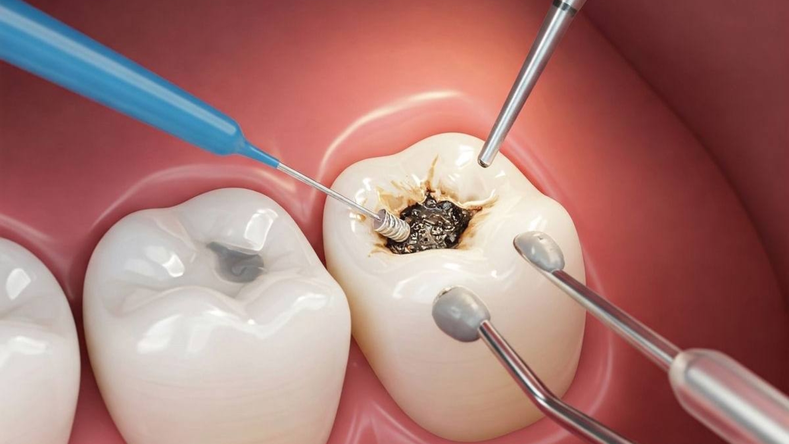
Early caries begins at a stage when the tooth surface may still appear intact and the patient experiences no pain, sensitivity, or other noticeable discomfort. Professor Alexander Von Breuer analyzes this phase as a critically important window for diagnosis because the demineralization process has already begun while a visible cavitated lesion may still be absent. At DentalClinic24, we evaluate early carious lesions not as insignificant cosmetic changes but as active biological processes capable of progressing from reversible enamel demineralization to irreversible destruction of hard dental tissues. Our objective is to determine lesion activity, identify the underlying causes of mineral loss, and select a treatment strategy that preserves the greatest possible amount of natural enamel without unnecessary mechanical preparation of the tooth.
The development of early caries results from a prolonged imbalance between enamel demineralization and its natural remineralization capacity. After the consumption of fermentable carbohydrates, bacteria within the dental biofilm metabolize sugars and produce organic acids. The acidity at the tooth surface increases, calcium and phosphate ions begin leaving the crystalline enamel structure, and the subsurface enamel gradually becomes porous. Saliva normally neutralizes acids and restores essential minerals, but frequent snacking, inadequate plaque removal, reduced salivary flow, or diminished buffering capacity prevent these natural repair mechanisms from compensating for ongoing damage. As a result, a chalky white spot develops, often remaining almost invisible under normal lighting conditions, particularly when located between adjacent teeth, within deep fissures, or alongside the margins of existing restorations.
At DentalClinic24, diagnosis extends far beyond searching for dark discolorations because color alone does not accurately indicate either the depth or biological activity of a carious lesion. The clinician carefully evaluates enamel gloss, translucency, surface texture, structural integrity, lesion location, plaque accumulation, and the condition of the surrounding gingival tissues. Active lesions typically present with a matte, rough appearance, whereas arrested lesions often become smoother and regain a more glossy surface. Following professional plaque removal and controlled air drying, subtle enamel changes that remain hidden under normal moisture conditions become clearly visible. Magnification, specialized illumination, clinical photography, and optical diagnostic technologies further assist in identifying alterations in enamel light transmission before obvious structural defects develop.
Interproximal early caries presents a particularly challenging diagnostic situation because these lesions develop between neighboring teeth where direct visual examination is impossible. Such lesions frequently remain undetected until they extend into dentin. Bitewing radiographs provide valuable information regarding the location and approximate depth of proximal demineralization. Nevertheless, radiographic findings should never be interpreted in isolation because very early lesions may remain radiographically undetectable, while the extent of radiographic shadowing does not always correspond precisely with biological lesion activity. Decisions regarding observation, remineralization therapy, or restorative intervention are made only after integrating all available clinical information, including surface characteristics, oral hygiene quality, individual caries risk, and changes documented during previous examinations.
At DentalClinic24, identifying the factors responsible for ongoing demineralization represents an essential part of every treatment plan because localized management alone cannot provide lasting clinical stability if the underlying causes remain unchanged. The progression of early caries is influenced by dietary sugar frequency, inadequate interdental cleaning, dental crowding, orthodontic appliances, insufficient fluoride exposure, reduced salivary flow, and anatomical areas where biofilm consistently accumulates. In orthodontic patients, early lesions frequently develop around brackets and attachments, while tightly positioned teeth often create inaccessible areas beyond the reach of conventional toothbrushes. Individual preventive programs therefore include personalized oral hygiene instruction, appropriate interdental cleaning devices, dietary counseling, and professionally selected remineralizing agents designed to strengthen enamel.
Treatment of early caries depends primarily on whether the external enamel surface remains intact and how biologically active the lesion appears. When no cavitation is present, conservative approaches aimed at strengthening enamel and reducing bacterial activity receive priority. Fluoride based formulations increase enamel resistance to acid attack while promoting the incorporation of essential minerals into its crystalline structure. Calcium and phosphate containing products may provide additional support for remineralization. In selected clinical situations, resin infiltration techniques are performed to penetrate porous enamel with a low viscosity material that blocks further acid diffusion while simultaneously reducing the visual appearance of white spot lesions. The choice of treatment depends on lesion depth, anatomical location, and the likelihood of continued progression.
Monitoring treatment outcomes is equally as important as initiating therapy. The clinician must confirm that the lesion surface has become denser, regained natural luster, and remains stable without enlargement over time. Follow up clinical photography, visual examinations, and radiographic monitoring when indicated provide objective documentation of treatment success. The absence of pain cannot be considered evidence of stability because early caries rarely produces subjective symptoms either before or after treatment. True clinical success is measured by biological arrest of the lesion, improved oral hygiene parameters, and a reduction in the development of new carious sites. If the lesion continues progressing or the enamel surface loses its integrity, the treatment plan is revised toward minimally invasive restorative management.
At DentalClinic24, we strive to distinguish lesions that genuinely require operative intervention from those capable of stabilizing through conservative management. Premature removal of healthy tooth structure results in irreversible enamel loss and initiates a restorative cycle in which each replacement restoration often requires progressively greater removal of natural tissue. Conversely, passive observation without proper risk assessment may allow the lesion to advance into dentin. Sound clinical decision making lies between these two extremes and is based on lesion activity, surface integrity, accessibility for oral hygiene, and patient compliance. This balanced philosophy enables us to treat the biological disease process rather than simply responding to its advanced structural consequences.
Early caries clearly demonstrates the importance of preventive dentistry before pain develops. Minimal enamel changes remain biologically reversible when detected promptly, interpreted accurately, and incorporated into a comprehensive preventive strategy. At Dental Clinic24, we combine advanced diagnostics, individualized risk assessment, biofilm control, and scientifically supported remineralization protocols to preserve natural tooth structure while avoiding unnecessary intervention. Early detection significantly reduces the likelihood of progression to deep caries, pulp inflammation, and extensive restorative treatment. Tooth preservation begins long before significant destruction occurs, at the moment when the clinician recognizes the earliest microscopic changes and makes a carefully reasoned clinical decision.
Previously, we wrote about Occlusal Wear Facets: How Specialists at DentalClinic24 Use Contact Marks to Diagnose Overloads