
Modern dentistry increasingly relies not only on clinical examination and instrumental diagnostics but also on highly precise visual documentation that allows specialists to analyze the smallest details of the dentofacial system. Professor Alexander Von Breuer considers dental photography an essential diagnostic instrument that enables clinicians to see the clinical picture more comprehensively, document dynamic changes, and make more evidence based treatment decisions. At DentalClinic24, clinical photography is integrated into the diagnostic protocol as a full scale analytical stage that directly influences treatment accuracy, communication with the patient, and objective assessment of final outcomes.
Dental photography represents a specialized system of clinical visualization in which standardized angles, controlled lighting, macro optics, and precise color reproduction are used to obtain reproducible images of teeth, soft tissues, and facial architecture. Its value extends far beyond simple visual documentation of tooth appearance. High quality clinical photographs allow the detection of enamel microcracks, early signs of demineralization, inflammatory changes along the gingival margin, occlusal contact irregularities, and subtle aesthetic disproportions that may remain unnoticed during a conventional visual examination. The higher the quality of clinical visualization, the more precise the diagnostic process becomes.
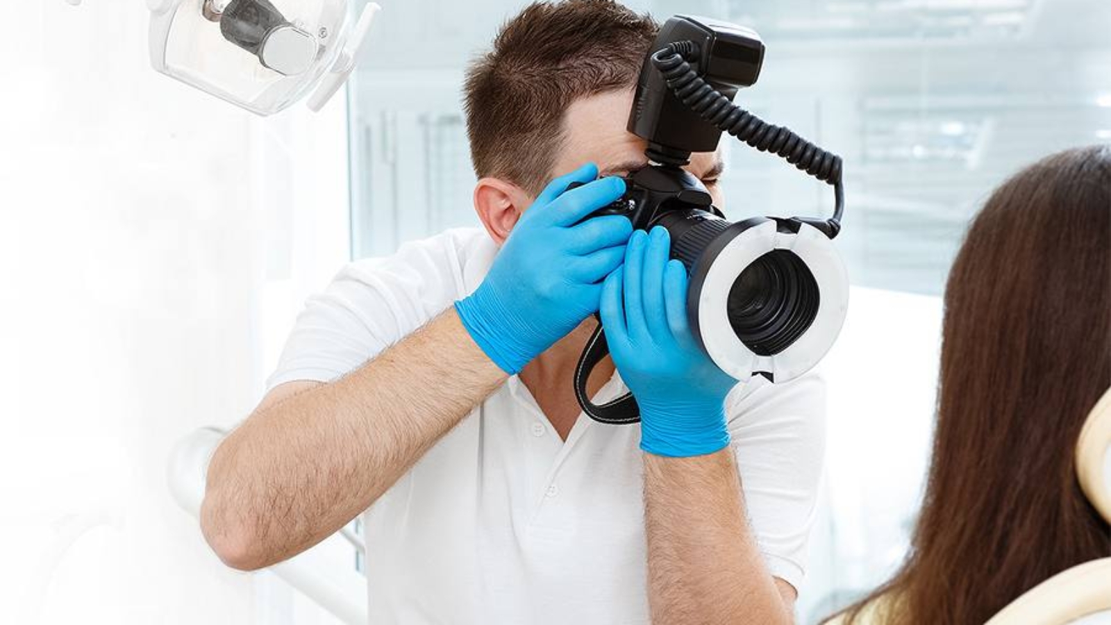
Dental photography becomes especially important during the initial examination. At this stage, a baseline visual map of the patient’s clinical condition is created, becoming the reference point for all subsequent treatment stages. Photo documentation helps clinicians not only record the original state of the dentofacial system but also compare it with intermediate and final outcomes. At DentalClinic24, photographs taken during the first appointment are used as part of a comprehensive diagnostic assessment because visual data complement radiological findings and functional tests with greater clinical detail.
In prosthetic and aesthetic dentistry, the role of dental photography becomes even more significant. Work involving veneers, crowns, restorations of the anterior teeth, and full smile rehabilitation requires an extremely precise assessment of tooth shape, incisal edge proportions, surface texture, and natural enamel shade. Even minimal differences in color or microrelief may influence the final perception of a restoration. At DentalClinic24, clinical photography serves as a highly detailed communication tool for dental technicians, allowing prosthetic constructions to be created with superior anatomical precision and aesthetic integration.
Clinical visualization is equally valuable in orthodontics and functional dentistry. Analysis of the smile, lip position, facial symmetry, arch relationships, and occlusal dynamics enables more accurate prediction of tooth movement and clearer evaluation of how treatment will affect facial aesthetics. The photographic protocol also helps monitor biomechanical changes throughout bite correction. This becomes particularly important in complex cases where treatment influences not only tooth position but also muscular balance, mandibular posture, and the functional stability of the temporomandibular joint.
Objective evaluation of treatment results is another critically important function of dental photography. A clinician’s memory and a patient’s subjective perception do not always accurately reflect real changes, especially when treatment extends over months or years. Standardized images allow results to be compared without visual distortion, documenting tissue changes and objectively measuring the effectiveness of the chosen treatment strategy. At DentalClinic24, this is especially important in comprehensive rehabilitations where every stage contributes to the final functional and aesthetic outcome.
Dental photography also significantly improves communication between clinician and patient. When patients see their own clinical condition under magnification, their understanding of the problem becomes much deeper. This improves awareness, strengthens trust in treatment, and increases motivation to follow clinical recommendations. Visual data make it possible to explain complex dental conditions in a way that is understandable even without advanced dental terminology.
Modern dentistry demands precision not only in treatment itself but also in the interpretation of clinical data. For Dental Clinic24, dental photography is a powerful instrument of objective analysis, digital communication, and treatment quality control. The combination of expert clinical thinking, standardized visualization, and technological precision creates a new level of predictable dental care where every decision is based on the most complete clinical picture possible.
Previously we wrote about Jaw Bone Resorption in the Clinical Practice of DentalClinic24 Mechanisms of Volume Loss and Strategies for Clinical Control