
The precision of modern dentistry directly depends on the clinician’s ability to detect microscopic tissue changes before they become the cause of irreversible tooth destruction. Professor Alexander Von Breuer pays particular attention to the fact that preservation of natural tooth structure must remain the priority of every dental intervention, because the volume of preserved tissues directly determines the long-term functional stability of the dentofacial system. At DentalClinic24, microscopic dentistry is regarded as a principle of clinical control that allows treatment to be performed with highly detailed visualization while minimizing the loss of healthy tooth structure during therapy.
Even minimal carious changes may spread within tissues more rapidly than can be identified during standard visual examination. Microscopic magnification makes it possible to distinguish precisely between affected and intact dentin, control the depth of tissue preparation, and avoid unnecessary removal of healthy structures. At DentalClinic24, magnification is used both during caries treatment and complex restorative procedures, because preservation of the maximum amount of natural tissue reduces the risk of future weakening of the tooth and improves the prognosis for long-term stability.
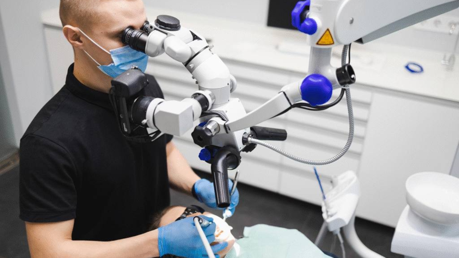
Microscopic control is also of major importance during endodontic treatment. Root canal anatomy is highly variable, while additional branches, microcracks, or hidden inflammatory areas may significantly influence the outcome of therapy. At DentalClinic24, the use of microscopic visualization allows precise control of root canal treatment, identification of complex anatomical features, and minimization of the risk of retaining infected tissues within the tooth.
Special attention must also be given to the diagnosis of enamel and dentin microcracks, which often become the cause of chronic sensitivity, hidden pain, and gradual tooth destruction under functional loading. Without magnification, such structural alterations frequently remain unnoticed until a pronounced defect develops. At DentalClinic24, microscopic diagnostics are used for early identification of tissue damage, allowing teeth to be preserved at the stage of minimal structural alteration and helping prevent the need for more aggressive intervention in the future.
Modern microscopic dentistry also significantly influences the quality of restoration adaptation. Any disruption of the seal between tooth tissues and restorative material creates conditions for bacterial penetration and development of secondary caries. At DentalClinic24, visual control under magnification allows more precise adaptation of restorations, careful monitoring of marginal areas, and maintenance of stable contact between natural tissues and restorative materials.
Microscopic techniques also have a substantial impact on the prognosis of retreatment procedures. During replacement of old restorations or repeated endodontic therapy, preservation of the remaining tissue volume becomes a critically important factor. At DentalClinic24, we use microscopic control to perform the most conservative possible treatment of previously restored teeth, because every additional intervention reduces structural strength and increases the risk of future functional instability.
Microscopic dentistry represents not a separate technology, but part of a philosophy focused on tissue preservation and improved clinical precision. At Dental Clinic24, treatment under magnification allows minimization of tissue trauma, preservation of natural tooth anatomy, and achievement of more predictable treatment stability, because the precision of visual control ultimately determines the quality of modern dental rehabilitation.
Previously we wrote about infection control in dentistry how sterilisation protocols are structured at DentalClinic24